

Volume 27 Number 1
High-risk foot care in private practice
Sarah Coombes
Keywords Diabetes, Multidisciplinary care, high risk foot
For referencing Coombes S. High-risk foot care in private practice. WP&R Journal 2019; 27(1):36-41.
DOI https://doi.org/10.33235/wpr.27.1.36-41
Abstract
Many rural areas of New South Wales, Australia, have limited access to community podiatry and no high-risk foot (HRF) clinics. Private practitioners in these areas develop their own multidisciplinary teams (MDT) involving local professionals of different disciplines to provide evidence-based care for their patients. This case study shows co-ordinated care in a rural setting for a disunited navicular fracture in a patient with diabetes T2, familial neuropathy, a history of osteomyelitis and previous minor foot amputations. It explores the difficulties of wound care when treating complex cases without access to immediate consultations across all the specialties and highlights the need for better access to MDT HRF clinics for those clients with complex care needs.
Introduction
The principle of diabetic foot ulcer care is to ensure optimal diabetes control, adequate blood supply, offload pressure from area, debride non-viable tissue and manage infection1. Patient-centred care can increase patient empowerment, reduce stress and improve outcomes in diabetes management2. Multidisciplinary team (MDT) care is preferred to allow for good communication and data collection amongst all professionals involved in the patient’s care3-6. Interdisciplinary care teams in dedicated high-risk foot (HRF) services have been shown to reduce hospital admission rates and improved patient outcomes for diabetic foot problems7.
This case study was done over a 15-month period from March 2017 to November 2018. Patient M is a 72-year-old female with idiopathic familiar axonal neuropathy and T2DM with peripheral neuropathy. M resides in a rural community 35 kilometres from a regional centre with both private and public hospitals. Extended informed consent, obtained both verbally and written, was given by the patient at the start of this study.
M received care from a team of specialists for her co-morbidities including vascular surgeon, neurologist, endocrinologist, physiotherapist, exercise physiologist and home care nursing. She had also seen an orthopaedic surgeon in previous years to consider surgery for foot deformity prior to amputations. This study describes her HRF management, focussing on the progressive deterioration of both feet from dactylitis to ulceration of one foot and fracture to below-knee amputation of the other.
Case study
M presented in March 2017 with pain in her left posterior-lateral heel and a wound on the apex of the third toe. Over a 12-month period, M’s foot conditions progressively deteriorated and she presented with multiple small wounds before an episode of dactylitis led to a chronic ulcer on her right first toe and she sustained a disunited navicular fracture on her left foot.
A care plan for M included regular podiatry review at six-week intervals, or earlier, if any problems. Her husband was happy to check her feet every day and actively helped with her foot care, including applying moisturiser daily. Home care nurses would help with dressings as needed. Her initial concerns were pain in her left heel and a University of Texas grade (UT) 1a ulcer8 on the apex of the right third toe caused by impact injury. On examination, her left heel had no oedema or erythema, and x-ray and ultrasound investigations were normal. Pain settled over the time taken for radiology tests with no intervention and was thought to be related to her neuropathy. The right toe wound healed over a four-week period using evidence-based (EB) wound management. In late November 2017, hyperkeratosis (a callous) developed on the dorsal interphalangeal-joint (IPJ) of her right second toe. Three weeks later M had developed a “sausage toe” (dactylitis) due to erythema and oedema, indicating early signs of osteomyelitis (OM)9. M was referred to her general practitioner (GP) for antibiotics10 and was subsequently referred to a local vascular surgeon for review. The vascular surgeon elected to amputate the toe and the pathology confirmed OM.
Within one month of the amputation, a callous developed on the apex of her right first toe. Due to M’s recent ulceration, amputation and OM, her care plan was reviewed. M agreed to podiatry visits four-weekly, increasing when required for wound care and/or callous debridement11. M admitted to finding more than two medical appointments per week exhausting. She was strongly encouraged to try and maintain all follow-ups with her health professionals within the recommended time to allow for early prophylactic care.
M has severe familial and diabetic peripheral neuropathy with multiple co-morbidities including lesser toe amputations and ulceration over a 10-year period prior to her initial visit with me. She appeared overwhelmed and depressed by her neuropathic lower limb pain, which escalated with her anxiety. She appeared to be happy in her relationship with her husband and liked to attend social events when possible.
Initial assessment findings
M presented with complex care needs due to neuropathy, diabetes and foot deformity. Reduced sensory nerve potential and conduction is common to both axonal and peripheral neuropathy12 and causes motor paresis and atrophy of small foot muscles contributing to foot deformity12 M had difficulty walking and used either a walking stick or wheelie walker, depending on distance. She had abducted feet, genu valgum and collapsing medial arches. She had first and second left toe amputations, with her remaining digits clawed and fixed. There was minimal callous on plantar pressure areas of her feet and remaining toes. She had well-fitted medical grade footwear (MGF) that supported her foot in gait with custom deflective insoles. Unfortunately, M disliked wearing these shoes.
Foot pulses were not palpable, but Doppler studies revealed biphasic pedal pulses indicating adequate blood flow13. A recent duplex ultrasound with the vascular surgeon had been reported unremarkable.
Evaluation of sensation using 10 g and 75 g monofilament fibres, vibration with 128 Hz tuning fork, hot/cold sensation and ankle reflex confirmed neuropathy12. M reported severe shooting pains in both thighs, the left worse than the right, by early afternoon that made moving very difficult. She reported an inability to keep her legs still at night due to severe pain in her thigh and calf, despite high levels of pain medication. These symptoms are consistent with both axonal and peripheral neuropathy12.
M’s BMI was above normal range. She ate fresh fruit and vegetables daily, but drank minimal water. She reported low levels of vitamins B12 and D, and magnesium, which may affect wound healing14.
Management and wound care
M’s right first toe ulcer developed due to friction when wearing her non-MGF footwear after amputation of the right second toe. Change in foot shape and badly fitting shoes caused a callous on the apex of the right first toe. Late presentation to the clinic for treatment resulted in the callous tearing off, leaving a skin tear injury. The wound degenerated over a three-week period from a category 2a skin tear15, to a 1 cm diameter UT1b8 ulcer with a friable hyper-granulating base and firm, blanched, sloping edges, despite EB wound management.
Ideally, M would have received treatment and assessment at an accredited MDT HRF clinic which can provide on-site access to imaging, pathology, and a foot specialist team to lead case conferencing and provide immediate therapy and referrals7,16. Immediate access to offloading boots and/or total contact casts (TCC) and loan of mobility equipment such as wheelchairs would also be available at no cost to the patient. The closest accredited HRF clinic is more than 300 kilometres away. This would have had a negative socio-economic impact on M due to accommodation costs and lack of social support, and possibly increase her already high anxiety. She found travel of 35 kilometres from home to our regional centre was both painful and tiring and did not want to travel any further than she had to for services.
As there was no HRF clinic nearby, M elected to have podiatry services in her local town in private practice. At every visit, wound characteristics, peri-wound skin, and wound measurements were assessed, wound dressing choices were evaluated, footwear and offloading devices were inspected and treatment adjustments were made accordingly. While the primary aim is to heal tissues, a history of amputations and OM indicates an ongoing high risk of recurrence17. An electronic patient record was utilised in M’s care, which facilitated accurate reporting and continuity of care3. Copies of these notes were sent to all team members by fax/email on the same day. M’s GP was informed of her progress after each visit and potential changes to medication, diet, social and psychiatric support suggested as appropriate.
Wound care
6 March 2018
M presented with a callous on the apex of her right first toe, which had dried causing the edges to lift and tear on bedclothes, leaving a category 2a skin tear15. The wound was 1 cm diameter with no exudate and no signs of infection. Treatment was conservative sharp wound debridement (CSWD) of residual callous on periwound and a foam dressing cap was applied to the whole toe for moisture balance and protection. The home nursing service was requested to redress and monitor. M was asked to present to the clinic if any deterioration was noted. A review was planned with the podiatrist for 14 days later.
20 March 2018
The right first toe UT1 ulcer was 1 cm diameter with a friable, hypergranulating wound bed, indicating localised infection10. The wound edges were firm, blanched, sloping and not undermining. There were no problems found with her MGF shoe fit. M had no fevers or chills and felt well. The ulcer was cleaned with normal saline and a dialkyl carbamoyl chloride Sorbact foam dressing made into a toe cap was applied. Due to the recent amputation of the adjacent toe from OM and ongoing oral antibiotics, an antimicrobial dressing was used prophylactically. Antibiotics are used up to 90 days post-surgery for OM and there is potential for reinfection in another wound within six weeks of previous surgery18. Reducing the superficial wound bioburden may help reduce that potential18. Sorbact binds bacteria and fungi into the dressing surface, rendering them inactive19. Foam is non-adhesive, protective, absorbs exudate away from the wound bed and potentiates moist wound healing10. Sorbact does not adhere to the wound surface, protects the wound with cushioning and does not add chemicals to the wound10. X-rays ordered prior to this appointment for the first toe views were unremarkable.
27 March 2018
The right first toe UT1b ulcer showed no change and treatment of CSWD and wound dressing regimen was repeated (Figure 1).
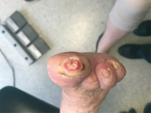
Figure 1: Wound on apex of right first toe.
Review with vascular surgeon 28 March 2018
The vascular surgeon requested conservative care with silver-impregnated foam dressings to be provided by the home nursing service twice-weekly and prescribed long-term Keflex 500 mg b.d. The nursing service requested podiatry review for CSWD, as needed.
M was reviewed at two-week intervals for CSWD. The right first toe UT1a ulcer had reduced in size to 0.4 mm diameter on 17 April 2018 and some granulation was noted in the wound bed. The nursing service continued care.
18 May 2018
M presented with a hot, swollen, painful left ankle, stating her “foot feels like it is falling off her leg”. She had marked collapse of the medial longitudinal arch and difficulty walking due the position of her foot (Figure 2). There was no history of falls or injuries to the area, M’s temperature was reported normal that morning at her GP visit, she had no fevers or chills but had a pain score of 5/10 on the verbal rating scale in her left ankle area.
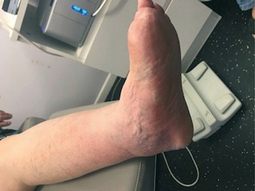
Figure 2: Initial presenting left foot deformity.
Right first toe
The UT1b ulcer measured 1.5 cm diameter and had deteriorated with undermined edges, heavy periwound callous with maceration, soft, fibrous slough over the wound bed and a mild odour. The wound probed but not to the bone. Due to the musty odour, indicating a high bioburden, the wound was cleaned with Prontosan wound irrigation solution for immediate reduction in contaminants10. As previous silver dressings had been effective in reducing the size of the ulcer a silver hydrofibre (Aquacel Ag) dressing was applied to autolytically debride slough and manage bioburden10. A Biatain foam toe cap was applied as a secondary dressing to facilitate moisture balance and offload pressure10.
To offload the acute left foot deformity M was strongly encouraged not to weight-bear while further investigations could take place1.
22 May 2018
X-rays showed a left foot disunited navicular fracture and mild bone pitting of the medial distal phalanx on the right foot. The right first toe wound bed was effectively debrided by Aquacel Ag. The UT1a ulcer measured 1.5 cm diameter, and wound bed was clean, with 25% granulation tissue. The edges were not undermining and there was no odour. The wound was cleaned with normal saline and the foam cap was applied. An antimicrobial dressing was not used as M was on long-term Keflex 500 mg b.d. and the wound bed was clean and granulating.
An urgent review was requested with the orthopaedic surgeon for the navicular fracture. M was advised to wear her own controlled ankle movement (CAM) boot, keep off her feet and check her left foot skin twice-daily.
23 May 2018
M continued to weight-bear with her CAM boot and within 24 hours she developed a category 2b skin tear 5 cm x 3 cm over the left medial navicular bulge from the CAM boot (Figure 3). She had not removed the boot as she felt more stable walking in it. The wound bed was oozing serous exudate with bruising in the deeper tissues. It was dressed with non-adherent foam to maintain moisture balance and provide protection, plus Zetuvit (combine) dressing for added protection10. Both dressings were adhered with Hyperfix tape individually to prevent inner dressing shearing under the top dressing. The dressing was covered with a Tubigrip stocking from toes to below knee to support the tissue, improve circulation and reduce oedema20. The CAM boot was modified to remove pressure on the deformity. M was encouraged to remain non-weight-bearing but she admitted to being frustrated by relying on her husband for all her needs and preferred to move around the unit herself as much as possible rather than wait for a wheelchair for toileting and so on. The orthopaedic surgeon was notified of the deterioration and an appointment was made with him for the following day (Figure 3).
There was no strikethrough on the dressing on the right first toe, so the dressing was left intact.
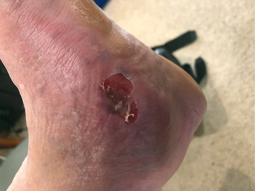
Figure 3: Skin tear after 24 hours of wearing CAM boot.
24 May 2018
The orthopaedic surgeon ordered a Charcot Restraint Orthotic Walker (CROW) as there are no technicians capable of managing a TCC in our area. The CROW cost M about $800 and took four days to manufacture. Costs for M were recouped by her NDIS care package. In an HRF clinic a TCC could have been applied immediately and would have been available through hospital funding.
The left foot wound was showing 50% strikethrough on the dressing. The wound was cleaned with normal saline and the foam/Zetuvit dressing was replaced. There was no strikethrough on the dressing on the right first toe so the dressing was left intact.
25 May 2018
The left foot deformity was increased and a UT1b ulcer had developed over the site of initial bruising. The ulcer measured 4 cm x 3 cm, within a category 2b skin tear area of 7 cm x 5 cm. The wound bed was blanched, with approximately 20% granulation tissue noted in small spots across the ulcer. The wound exudate was haemoserous and moderate. The ankle was hot and swollen with inflammation from the increasing collapse of the foot. Sorbact foam was used for its antimicrobial properties with an overlying Zetuvit pad for protection. The wound was cleaned with normal saline and non-viable skin tear tissue was debrided. M had no fevers or chills and was still on antibiotics.
The right toe dressing regimen was maintained.
29 May 2018
The prosthetist requested a less bulky dressing due to limited space within the CROW boot. When using a TCC, the size of the dressing is incorporated into the device. A CROW boot is useful to support the collapsing arch but did not allow changes in leg size due to the reduction in oedema. A TCC can be removed and replaced to accommodate leg size as required.
The left foot UT1b wound measured 4 cm x 2 cm with a central deeper crater measuring 2.5 cm x 2 cm. Inflammatory heat from the ankle was drying the ulcer bed. The wound was cleaned with normal saline. Due to the dry wound bed Flaminal hydrogel — an enzyme alginogel that promotes autolytic debridement, maintains moisture balance, protects wound edges and epithelial cells and reduces microbial burden — was applied10. A silicone foam secondary dressing (Mepilex Border) was applied for protection and moisture balance. Silicone foam is resilient but less bulky when compared with combine dressings, allowing for good boot fit. The dressing was fixed with Hyperfix tape around the borders to prevent shearing of the dressing if the adhesive failed to grip.
The right first toe UT1b ulcer was extending dorsally and a small sinus noted but there was 30% granulation tissue in the wound bed. The periwound callous was minimal and the wound bed was shallow and beginning to dry. The ulcer was dressed with Flaminal hydro gel for moisture balance, autolytic debriding and antimicrobial properties10 with a foam toe cap.
June 2018
M was due to review in two days but had to extend to four days due to four other medical appointments. At her follow-up podiatry appointment the head of the talus had broken through the medial side of her foot (Figure 4). The orthopaedic surgeon was notified and she was immediately taken to the private hospital by her husband. Within hours of admission she began to develop sepsis and within 12 hours was transferred to the public hospital due to a heart attack. At this point the vascular and orthopaedic surgeons felt that M’s life was at risk without immediate amputation of the left leg below the knee and surgery was completed within 48 hours.
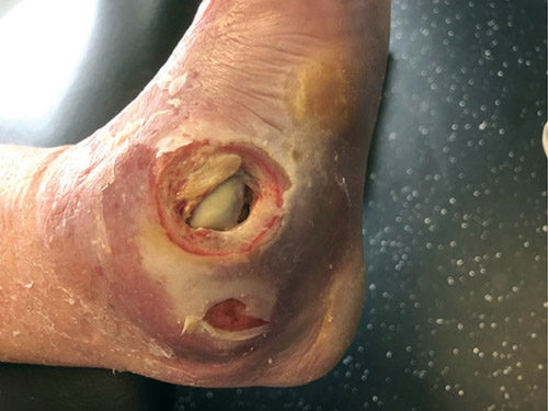
Figure 4: Breakthrough of talus at medial left foot ankle.
November 2018
M’s recovery and rehabilitation has been slow but steady and she presented in early November with a prosthetic leg, which she was managing well. Due to the enforced limited weight-bearing, the right first toe ulcer is now closed but fragile and M now wears her MGF shoes all the time to give her stability due to the prosthesis.
Discussion
Collaborative MDTs are considered to be the gold standard in diabetic HRF care7,21. In private practice an MDT can be created as needed by referring to the local specialist with the GP as team leader. Interprofessional relationships are critical to HRF care as a private practitioner21.
At initial presentation, M appeared exhausted by her foot problems. She had been unable to attend podiatry clinics regularly due to her ill health or other delays, meaning she missed her appointment and could not reschedule for two–three weeks due to practitioner bookings. She appeared to be depressed but had no formal diagnosis. Depression and neuropathy may be bi-directional, with one increasing symptoms of the other and vice versa22 and depression may reduce capability of compliance with medications, exercise, diet and follow-up care instructions22. Her initial appointment ran well over the allocated time due to discussion about her foot problems and helping her to determine a care pathway she felt she could achieve in relation to follow-up appointments and home monitoring. Lengthy appointments may cause anxiety in patients in a private practice where the fees are related to the time spent in consultation. This may lead to the client cutting the appointment and discussion short in order to ‘save money’ and important instructions and explanations may not be absorbed by the patient. Despite agreement, M continued to extend gaps between foot appointments due to other medical reviews. Appointments at an HRF clinic could have reduced the number of appointments she had to attend, and/or reduced travel between specialists by allocating appointments in the same venue, which may have improved her outcomes23. Lengthy appointment times with no financial penalty may have resulted in better understanding of the critical nature of her foot problems and the need for excellent adherence to non-weight bearing and skin inspection instructions.
Higher rates of lower limb amputations are seen in regional and remote areas when compared with large city areas7. Access to critical care services for regional and remote patients can be costly, both financially and emotionally if they are required to travel to major centres for treatment. Access to specialist care is limited by the number of specialists available in the region and can be severely delayed due to periodic leave or high emergency case loads. The new accredited HRF service minimum standards requirement for staffing in rural and remote areas is: an advanced scope of practice nurse practitioner with access to a consulting physician, a credentialled diabetes educator and a senior podiatrist7,p.13. Access to both vascular and orthopaedic surgeons is also required7,p.13. Where this level of staffing is not possible, utilising technology for video conferencing may help to provide better critical HRF care to regional and remote clients in their own areas24.
Over the past year, I have had four clients with complex HRF conditions in my private practice. It has highlighted to me that the establishment of medical-led accredited HRF clinics in rural and regional areas is critical to provide excellence in MDT lower limb wound care.
Conflict of Interest
The author declares no conflict of interest.
Funding
The author received no funding for this study.
Author(s)
Sarah Coombes
MHSc(Pod)
Twinkletoes Podiatry Wauchope NSW
Email twinklespod@gmail.com
References
- International Best Practice Guidelines: Wound Management in Diabetic Foot Ulcers. Wounds International; 2013. Available from: www. woundsinternational.com World Union of Wound Healing Societies (WUWHS).
- Delaney LJ. Patient-centred care as an approach to improving health care in Australia. University of Canberra, Australia; 2017. https://doi.org/10.1016/j.colegn.2017.02.005 1322-7696/Crown Copyright © 2017 Published by Elsevier Ltd on behalf of the Australian College of Nursing Ltd.
- Mitchell G, Brown R, Erikssen L, Tieman J. Multidisciplinary care planning in the primary care management of completed stroke: a systemic review. BMC Family Practice 2008;9(44). doi:10.1186/1471-2296-9-44.
- Considine R, Tozer J, Milne H, Chappe M. Evaluation of the Super clinics Program 2006–2007. Australian Government Department of Health; 2012. http://www.health.gov.au/internet/publications/publishing.nsf/Content/GPSuperClinicsEvaluation-toc~discussion~progresstowardsachieving~multidisciplinarycare
- Foster A. Changes in the care of the diabetic foot: part one. Practical Diabetes Int 2001;18(4):134–138.
- Foster A. Changes in the care of the diabetic foot: part two. Practical Diabetes Int 2001;18(5):165–169.
- The National Association of Diabetes Centres and the Australian Diabetes Society. 2018 NADC Collaborative Interdisciplinary Diabetes High Risk Foot Services (HRFS) Standards © National Association of Diabetes Centres Sydney Version 1.1 https://nadc.net.au/hrfs-accreditation/.
- Oyibo S, Jude E, Tarawneh I, Nguyen H, Harkless L, Boulton A. A comparison of two diabetic foot ulcer classification systems. The Wagner and the University of Texas wound classification systems. Diabetes Care 2001 Jan;24(1):84–88. doi.org/10.2337/diacare.24.1.84
- Dinc T, Kocaoglu H, Kayilioglu S, Duzgun A, Coskun, F. Sausage toe: an upsetting symptom in diabetic patients. Int J Diabetes Dev Ctries 2016;37(4).
- Wounds UK Best Practice Statement. The use of tropical antimicrobial agents in wound management, 3rd edn. London: Wounds UK; 2013.
- Boulton AJM, Armstrong DG, Albert SF et al. Comprehensive foot examination and risk assessment: a report of the task force of the Foot Care Interest Group of the American Diabetes Association, with endorsement by the American Association of Clinical Endocrinologists. Diabetes Care 2008;31(8):1679–85. doi: 10.2337/dc08-9021.
- Misra UK, Kalita J, Nair PP. Diagnostic approach to peripheral neuropathy. Ann Indian Acad Neurol 2008 Apr–Jun;11(2): 89–97.doi: 10.4103/0972-2327.41875.
- Vriens B, D’Abate F, Ozdemir B et al. Clinical examination and non-invasive screening tests in the diagnosis of peripheral artery disease in people with diabetes-related foot ulceration. Diabet Med 2018 Jul;35(7):895–902. doi: 10.1111/dme.13634.
- Hoffman K. When vitamin and nutritional deficiencies cause skin and nail changes. Podiatry Today 2015 Jan;28(1). https://www.podiatrytoday.com/when-vitamin-and-nutritional-deficiencies-cause-skin-and-nail-changes
- Carville K, Lewin G, Newall N, Haslehurst P, Michael R, Santamaria N, Roberts P. STAR: A consensus for skin tear classification. Primary Intention 2007;15(1):18–28.
- ACI NSW Agency for clinical Innovation. Standards for High Risk Foot services (HRFS) in NSW ACI Endocrine Network July 2014 version 11.
- Weledji E, Fokam P. Treatment of the diabetic foot — to amputate or not? BMC Surgery 2014;14(1). doi.org/10.1186/1471-2482-14-83
- Giurato L, Meloni M, Izzo V, Uccioli L. Osteomyelitis in diabetic foot: A comprehensive overview. World J Diabetes 2017 Apr 15;8(4):135–42. doi: 10.4239/wjd.v8.i4.135.
- Totty JP, Bua N, Smith GE et al. Dialkylcarbomoyl chloride (DACC)-coated dressings in the management and prevention of wound infection: a systemic review J. Wound Care Mar 2017 2;26(3). doi: 10.12968/jowc.23017.26.3.107.
- Thomas Hess C. Checklist for factors affecting wound healing. Adv Skin Wound Care April 2011;24(4):192. doi: 10.1097/01.ASW.0000396300.04173.ec.
- Department of Health, Western Australia. High Risk Foot Model of Care. Perth: Health Networks Branch, Department of Health, Western Australia; 2010.
- Bartoli F, Carrà G, Crocamo C et al. Association between depression and neuropathy in people with type 2 diabetes: a meta-analysis: Depression and neuropathy in type 2 diabetes. Int J Geriatr Psychiatry 2016 Jan;31(8):829–836.
- Morrice DJ, Bard JF, Koenigca KM. Designing and scheduling a multidisciplinary integrated practice unit for patient-centred care. Health Systems 2019;1–24. https://doi.org/10.1080/20476965.2019.1569481
- Smith-Strom H, Iversen MM, Graue M, Skeie, Kirkevold M. An integrated wound-care pathway, supported by telemedicine, and competent wound management — Essential in follow-up care of adults with diabetic foot ulcers Int J Med Inform 2016;94:59–66. http://dx.doi.org/10.1016/j.ijmedinf.2016.06.020 1386-5056

