Ahead of Print
Non-healing foot ulcers in diabetes mellitus and role of biopsy: a report of two cases
Giovanni de Gennaro, Walter Baronti, Laura Sambuco, Edoardo Trapani, Loredana Rizzo
Keywords diabetic foot ulcer; tissue biopsy; chronic wound; skin cancer; melanoma
For referencing de Gennaro G ,et al. Non-healing foot ulcers in diabetes mellitus and role of biopsy: a report of two cases. Journal of Wound Management. 2024;25(3):to be assigned.
DOI
10.35279/jowm2024.25.03.02
Submitted 10 February 2024
Accepted 4 June 2024
Abstract
Diabetic foot ulcers are a severe complication of diabetes mellitus and are commonly associated with peripheral artery disease and/or neuropathy. An inadequate treatment or the presence of concomitant infection may result in a delay in wound healing. However, in some circumstances, non-healing wounds may hide other underlying diseases, which in some cases may be malignant. A biopsy can be a useful tool to investigate chronic wounds to achieve early diagnosis with the aim of establishing an adequate treatment and to improve the prognosis. We present two clinical cases in which biopsy revealed the presence of malignancy in non-healing chronic ulcers.
Key messages
In diabetic patients, chronic foot ulcers may hide other underlying diseases, which in some cases may be malignant.
This paper describes two clinical cases in which skin cancer was misdiagnosed as diabetic foot ulcer.
Biopsy is crucial to exclude diseases other than diabetes that can be malignant. Early diagnosis is essential to establish adequate treatment and improve prognoses.
Introduction
Diabetic foot ulcers (DFU) are one of the most severe complications of diabetes mellitus and are associated with high morbidity and mortality rate, as well as placing a high economic burden on the healthcare system.1–3 More than a quarter of non-healing DFU may lead to lower extremity amputation within a period of 18 months after the wound’s first manifestation.4
Diabetic neuropathy and peripheral arterial disease (PAD) are usually the main causes of DFU.5 Wound healing delay may result from conditions such as: inadequate treatment, impaired arterial or venous circulation, concomitant infection, immunocompromised status, and older age.6 However, in some circumstances non-healing ulcers may hide other underlying diseases, which in some cases may be malignant.7 Thus, guidelines and experts recommend biopsying atypical chronic ulcers for differential diagnoses or inappropriate clinical progression.8 A biopsy can detect cancer and provide further information on the nature of the wound, including its potential for infection and its impediments to healing.9 We report here on two cases of diabetic patients presenting with chronic wounds poorly responsive to treatment, in which skin biopsy performance was crucial for an accurate diagnosis.
Case 1
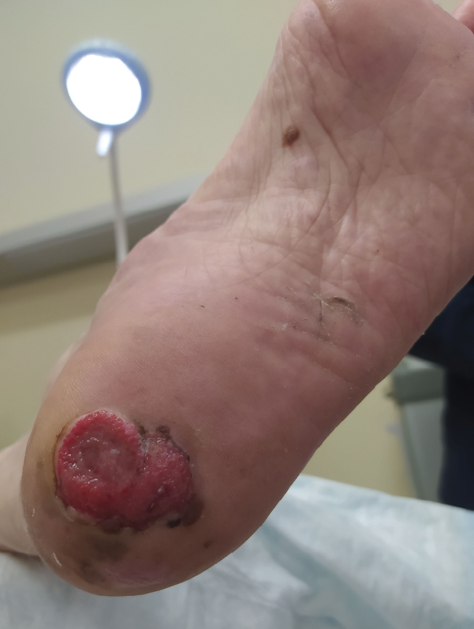
An 88-year-old Caucasian male patient with type 2 diabetes mellitus (T2DM) came to our Diabetic Foot Clinic for the evaluation of a non-healing ulcer involving the left plantar rear foot, present for more than two months (Figure 1).

Figure 1. Foot lesion in the first patient.
In addition to T2DM, his medical history included: hypertension, diabetic peripheral neuropathy (NP) and chronic obstructive pulmonary disease. The patient had no previous history of non-healing wounds, PAD, or of being immunocompromised.
The average wound area was 14cm2 by the manual planimetric method; the lesion was characterised by hyper-granulation tissue and hyper-pigmentation of the peri wound skin. No signs of local infection were present. At the beginning, the ulcer had been treated with dressings, topical antiseptic and local offloading, with no evidence of improvement.
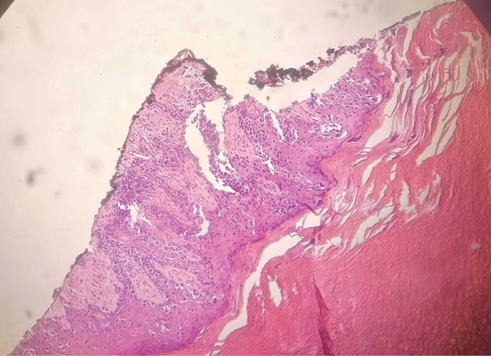
Due to the atypical appearance of the lesion, we decided to perform a biopsy, which was diagnostic for nodular melanoma (Figure 2). Disease staging confirmed the presence of metastases to the lungs and inguinal and hilar lymph nodes. In consideration of the patient’s age and comorbidities, no specific treatment was performed. The patient died six months after the diagnosis.

Figure 2. Histological pattern of nodular melanoma.
Case 2
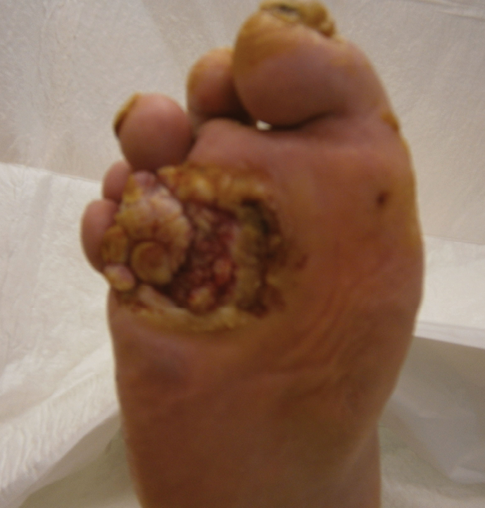
A 63-year-old Caucasian male came to our attention due to a non-healing plantar ulceration developed on his right forefoot (Figure 3). The patient had a seven-year history of T2DM, complicated by NP. There was no significant vascular defect; no history of trauma or previous DFU was reported. Medical history included hypertension, chronic renal disease, and previous myocardial infarction.

Figure 3. Foot lesion in the second patient.
The ulcer of 33cm2 was covered and surrounded by abundant hyperkeratosis; after keratolysis, inspection revealed the presence of exuberant granulation tissue with bleeding tendency. The wound was present for less than a year and had been treated with local dressing, rest, and an offloading device.
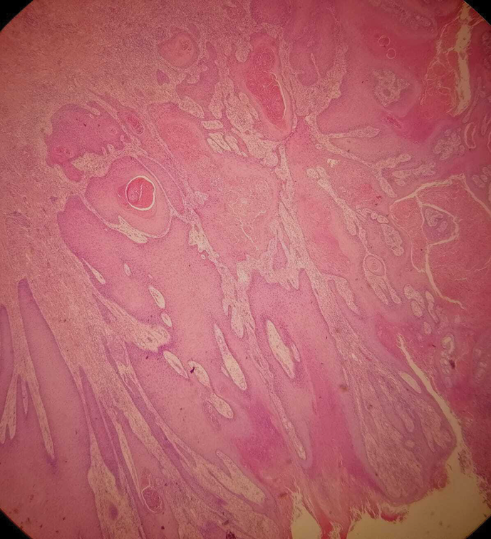
Given the chronic nature of the wound, an X-ray was performed and revealed osteomyelitis in the proximal end of the right fourth metatarsal bone. Furthermore, considering the unusual characteristics of the lesion, we also decided to perform a biopsy. In this case histopathology was remarkable for a well-differentiated squamous cell carcinoma (SCC), with an ulcerated pattern, infiltrating the skeletal muscle (Fig. 4). Lisfranc amputation procedure was performed. A computed tomography scan excluded metastatic spread of the disease. After the surgical wound healing process, the patient remained active and was able to walk by wearing proper footwear and a custom-made insole, without the need of further walking aids.

Figure 4. Histological pattern of squamous cell carcinoma.
Discussion
Melanoma and SCC are two of the most common skin cancers with high mortality rates worldwide.10,11 Among all melanomas, the nodular subtype has poorer prognoses,12 especially when arising on the foot, accounting for 35% of ultimately fatal cases of melanoma.13 Malignant melanoma has a high tendency to disseminate: after primary tumor excision about 30% of patients develop metastasis in various organs14; in the presence of nodal metastasis, the five-year survival rate decreases by approximately 50%.15 Nodular melanomas rarely occur on the foot; typically, they might seem symmetrical with well-defined borders or ill-defined, resembling a cauliflower. Their pigmentation ranges from black/brown to red/pink. The growth occurs vertically, progressing quickly and aggressively into deeper tissues. This is a specific characteristic for this subtype of melanoma.16 In the first case reported, the ulcer presented an exuberant granulation tissue with surrounding hyperpigmentation, similar characteristics to those just described. For this reason, we decided to perform a wound biopsy.
In the second case, the patient presented a non-healing plantar forefoot lesion covered and surrounded by abundant hyperkeratosis, resembling a neuropathic ulcer, but a biopsy revealed a high-risk SCC. The metastatic potential of a high-risk SCC is similar to that of melanoma17 and the annual disease-specific mortality is estimated to be 1.5–2.1%.18 Factors associated with local recurrence and metastases are: diameter >2cm, depth >2mm or beyond subcutaneous fat, perineural involvement, poor differentiation. Furthermore, SCC arising from a leg ulcer, burn scar and other chronic wounds have a reported metastatic risk of 26%.19 An uncommon type of well-differentiated SCC is verrucous carcinoma, a slow-growing lesion that does not frequently metastasize, although it is highly prone to recurrence and local invasion.20 Verrucous carcinoma, in its subtype called cuniculate epithelioma, is most commonly found on the weight-bearing surface of the foot and appears as an exophytic tumor, warty or cauliflower-like, with keratin-filled sinus tracts and malodorous exudate.21 The first-line treatment is surgical excision despite being particularly challenging due to the anatomic location and requirement for margin control while preserving healthy tissue, covering abnormalities, and maintaining acceptable function.20
Despite only few cases having been reported so far, SCC has been described as a complication of DFU22 or chronic osteomyelitis,23 and both patterns were detected in our patient.
Chronic ulcers and burn scars may be linked to SCC, which usually starts in the upper layers of the skin in areas continuously exposed to an inflammatory stimulus for an extended period,24,25 but no history of trauma was reported in our case. Chronic wounds that evolve in neoplasm are defined as Marjolin ulcers. The average latency period from the time of the initial triggering wound to the discovery of malignant degeneration is between 30 and 35 years,26 but also rare cases of acute Marjolin ulcer have been reported, occurring within 12 months of the ulcer development.27 In our patient the wound had been present for almost a year, which highlights how unusual the case was. Marjolin ulcers are often aggressive and carry a poor prognosis: metastases are found in up to 27% of cases and the recurrence rate after surgical resection is up to 50%.28 Melanoma and SCC are two malignant diseases, which can be misdiagnosed when present in the diabetic foot as they can mimic DFU, as neuropathic ulcers. Both patients had T2DM complicated by NP. Moreover, the ulcer site was in typical pressure point in both cases; in the second patient the ulcer was located on an area of hyperkeratosis, appearing as pressure ulcer. Therefore, both lesions received the same treatment as neuropathic ulcers, but, due to a lack of improvement, a skin biopsy was performed.
Clinicians should consider biopsy to rule out other ulcer causes if the ulcer does not heal following therapy and no cause for an intractable ulcer has been identified.8 The biopsy should be taken from the wound edge, including epidermidis, dermis and sufficient subcutaneous tissue. Among several types of skin biopsy techniques, the excisional is the preferred one when evaluating cutaneous tumors.29 However, there is no consensus regarding the appropriate timing for a wound biopsy in a chronic lesion. In general, a biopsy procedure should be conducted on wounds not responding to six or more weeks of standard treatment or continuously opened without signs of healing for three months.30 This procedure is crucial to exclude diseases other than diabetes that can sometimes be malignant. An early diagnosis could avoid systemic dissemination, limb loss, and death of the patient.
Conclusion
In conclusion, our cases demonstrate that lower extremity ulcers in diabetic patients may have an etiology other than diabetes. The medical history and the presence of atypical characteristics must suggest the execution of further diagnostic investigations, as biopsy, to exclude other diseases, which in some cases may be malignant. Therefore, early diagnosis is essential to establish adequate treatment and improve prognoses.
Future research
Further studies are needed to identify which characteristics are suspicious in chronic wounds and to establish the appropriate timing for skin biopsy performance.
Abbreviations
DFU: diabetic foot ulcers
PAD: peripheral arterial disease
T2DM: type 2 diabetes mellitus
NP: peripheral neuropathy
SCC: squamous cell carcinoma
Statement of ethics
All diagnostic and therapeutic procedures were in accordance with ethical standards of the institutional and national research committee and with the 1964 Helsinki Declaration and its later amendments or comparable ethical standards. Informed consent was obtained from the patients included in the study.
Conflict of interest
The authors have no conflicts of interest.
Funding
None declared.
Author(s)
Giovanni de Gennaro*, Walter Baronti, Laura Sambuco, Edoardo Trapani, Loredana Rizzo
Metabolic Diseases and Diabetes Unit, Misericordia Hospital, Grosseto, Italy
*Corresponding author email giovanni.degennaro@uslsudest.toscana.it
References
- Lazzarini PA, Pacella RE, Armstrong DG, van Netten JJ. Diabetes-related lower-extremity complications are a leading cause of the global burden of disability. Diabet Med. 2018;10.1111/dme.13680
- Jupiter DC, Thorud JC, Buckley CJ, Shibuya N. The impact of foot ulceration and amputation on mortality in diabetic patients. I: From ulceration to death, a systematic review. Int Wound J. 2016;13(5):892–903.
- Boulton AJ, Vileikyte L, Ragnarson-Tennvall G, Apelqvist J. The global burden of diabetic foot disease. Lancet. 2005;12;366(9498):1719–1724.
- Singh N, Armstrong DG, Lipsky BA. Preventing foot ulcers in patients with diabetes. JAMA. 2005;293(2):217–228.
- Hingorani A, LaMuraglia GM, Henke P, Meissner MH, Loretz L, Zinszer KM, et al. The management of diabetic foot: A clinical practice guideline by the Society for Vascular Surgery in collaboration with the American Podiatric Medical Association and the Society for Vascular Medicine. J Vasc Surg. 2016;63(2S):3s–21s.
- Sen CK, Gordillo GM, Roy S, Kirsner R, Lambert L, Hunt TK, et al. Human skin wounds: a major and snowballing threat to public health and the economy. Wound Repair Regen. 2009;17(6):763–771.
- Yang D, Morrison BD, Vandongen YK, Singh A, Stacey MC. Malignancy in chronic leg ulcers. Med J Aust. 1996;164(12):718–720.
- Isoherranen K, O’Brien JJ, Barker J, Dissemond J, Hafner J, Jemec GBE, et al. Atypical wounds. Best clinical practice and challenges. J Wound Care. 2019;28(S6):S1–s92.
- Senet P, Combemale P, Debure C, Baudot N, Machet L, Aout M, et al. Malignancy and chronic leg ulcers: the value of systematic wound biopsies: a prospective, multicenter, cross-sectional study. Arch Dermatol. 2012;148(6):704–708.
- Perez M, Abisaad JA, Rojas KD, Marchetti MA, Jaimes N. Skin cancer: Primary, secondary, and tertiary prevention. Part I. J Am Acad Dermatol. 2022;87(2):255–268.
- Rogers HW, Weinstock MA, Feldman SR, Coldiron BM. Incidence estimate of nonmelanoma skin cancer (Keratinocyte Carcinomas) in the U.S. population, 2012. JAMA Dermatol. 2015;151(10):1081–1086.
- Carrera C, Gual A, Díaz A, Puig-Butillé JA, Noguès S, Vilalta A, et al. Prognostic role of the histological subtype of melanoma on the hands and feet in Caucasians. Melanoma Res. 2017;27(4):315–320.
- Shaikh WR, Xiong M, Weinstock MA. The contribution of nodular subtype to melanoma mortality in the United States, 1978 to 2007. Arch Dermatol. 2012;148(1):30–36.
- Essner R, Lee JH, Wanek LA, Itakura H, Morton DL. Contemporary surgical treatment of advanced-stage melanoma. Arch Surg. 2004;139(9):961–966; discussion 6–7.
- Sanlorenzo M, Osella-Abate S, Ribero S, Marenco F, Nardò T, Fierro MT, et al. Melanoma of the lower extremities: foot site is an independent risk factor for clinical outcome. Int J Dermatol. 2015;54(9):1023–1029.
- Schade VL, Roukis TS, Homann JF, Brown T. “The malignant wart”: a review of primary nodular melanoma of the foot and report of two cases. J Foot Ankle Surg. 2010;49(3):263–273.
- Karia PS, Han J, Schmults CD. Cutaneous squamous cell carcinoma: estimated incidence of disease, nodal metastasis, and deaths from disease in the United States, 2012. J Am Acad Dermatol. 2013;68(6):957–966.
- Waldman A, Schmults C. Cutaneous Squamous Cell Carcinoma. Hematol Oncol Clin North Am. 2019;33(1):1–12.
- Rowe DE, Carroll RJ, Day CL, Jr. Prognostic factors for local recurrence, metastasis, and survival rates in squamous cell carcinoma of the skin, ear, and lip. Implications for treatment modality selection. J Am Acad Dermatol. 1992;26(6):976–990.
- Shwe Daniel S, Cox SV, Kraus CN, Elsensohn AN. Epithelioma Cuniculatum (Plantar Verrucous Carcinoma): a systematic review of treatment options. Cutis. 2023;111(2):e19-e24.
- Penera KE, Manji KA, Craig AB, Grootegoed RA, Leaming TR, Wirth GA. Atypical presentation of verrucous carcinoma: a case study and review of the literature. Foot Ankle Spec. 2013;6(4):318–322.
- Larijani B, Tavangar SM, Bandarian F. Squamous Cell Carcinoma arising in a chronic, nonhealing diabetic foot ulcer. Wounds. 2017;29(7):e48-e50.
- Saglik Y, Arikan M, Altay M, Yildiz Y. Squamous cell carcinoma arising in chronic osteomyelitis. Int Orthop. 2001;25(6):389–391.
- Tobin C, Sanger JR. Marjolin’s Ulcers: A case series and literature review. Wounds. 2014 Sep;26(8):248–254.
- Muzic JG, Schmitt AR, Wright AC, Alniemi DT, Zubair AS, Olazagasti Lourido JM, et al. Incidence and trends of basal cell carcinoma and cutaneous squamous cell carcinoma: a population-based study in Olmsted County, Minnesota, 2000 to 2010. Mayo Clin Proc. 2017;92(6):890–898.
- Pekarek B, Buck S, Osher L. A comprehensive review on Marjolin’s Ulcers: diagnosis and treatment. J Am Col Certif Wound Spec. 2011;3(3):60–64.
- Tisch Mendes J, Klein RJ. The value of a biopsy in chronic ulcers: a case series. Wounds. 2022;34(4):119–123.
- Shah M, Crane JS. Marjolin Ulcer [updated 2023 Jun 28] StatPearls. Treasure Island (FL). Available from: https://www.ncbi.nlm.nih.gov/books/NBK532861/
- Beard CJ, Ponnarasu S, Schmieder GJ. Excisional Biopsy [updated 2022 Aug 29] StatPearls. Treasure Island (FL). Available from: https://www.ncbi.nlm.nih.gov/books/NBK534835/
- Robson MC, Cooper DM, Aslam R, Gould LJ, Harding KG, Margolis DJ, et al. Guidelines for the treatment of venous ulcers. Wound Repair Regen. 2006;14(6):649–662.
