

Volume 25 Number 3
Care of oedematous skin in a resource-poor environment: a commentary of practice strategies to address a global community need
Terence J Ryan
Abstract
The emphasis of this paper is to discuss low-cost, self-help interventions for oedematous skin which can be utilised in low-resource communities or settings. In particular, the emerging understanding of the role of epidermal cytokines in repair, as a source of stimulating dermal oedema will be overviewed. Venous overload, due to gravitational effects, is a common contributor to lower limb oedema. A combination of epidermal cytokine activity and chronic ambulatory venous hypertension can overload a failing lymphatic system which, in the absence of overload, may not develop lymphoedema. The epidermis may switch off cytokine production when transepidermal water loss is reduced by appropriate washing and emollient application. Venous overload is helped by elevation and ankle movements, whilst breathing aids both lymphatic and venous drainage. These are low-cost self-help interventions which have been found to be helpful in resource-poor countries and can be recommended and promoted in the management of lymphoedema in a wide range of health care settings and environments.
Commentary Introduction
It has long been known that oedema delays healing1, but it has only recently been identified and emphasised that oedematous skin is not only unhealthy, but the epidermis is itself contributing to the oedema2-4. The major causes of oedema include cardiac failure, malignancy, venous hypertension or parasitic blockage of the blood or lymphatic vascular systems, and local inflammation. However, it is the response of the epidermis to oedema that partially explains lymphoedema and accounts for the benefits of meticulous skin care. The epidermis, when it is in good health, is an effective barrier, has sweat glands that contribute to thermoregulation, its sensory function is finely tuned without itch or pain and, by looking good, contributes to feeling good. When healthy, the skin has almost no ‘switching on’ of innate repair mechanisms, with only occasional mitotic activity. Most of the upper (outer) half of the epidermis, is anaerobic and the cells are tightly knit without nuclei.
In contrast, an unhealthy epidermis, such as in oedematous skin, is very different in both appearance and functionality. Oedematous skin initiates repair requirements with a ‘switched on’ repair mode including many mitotic figures and the dividing cells jostling for position which eventually exfoliate. The demand for oxygen will extend through most of the epidermis and often additional white cells will infiltrate further, increasing demands for oxygen. An active neutrophil can demand 50 times as much oxygen as any other skin cell. Importantly, the repair mode includes switching on the production of a range of inflammatory cytokines, many of which migrate into the dermis, where they greatly increase the permeability of the upper dermal capillary bed and stimulate the arrival of white cells2.
The majority of persons following a mastectomy, or infection from the mosquito-borne filariasis, do not develop lymphoedema until something else happens to overload the failing lymphatic system, including local trauma, bee sting or a bacterial cellulitis causing an inflammatory response. Appropriate management of fissures and interdigital crevasses has long received attention in the care of lymphoedema6,7 (Figure 1).
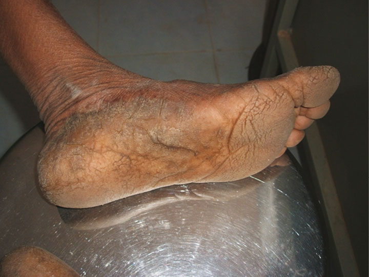
Figure 1: Fissures due to lack of moisturisation common in persons who do not wear shoes. They are entry points for soils irritants and bacteria.
Venous overload is another factor that contributes to lymphoedema, with gravity playing a role in overfilling the veins. Ambulatory venous hypertension is an underestimated inducer of lymphatic overload when the system is partially non-functioning and examination for dilated veins should always be conducted with the person in the upright position. Lymphoedema of the legs characteristically manifests as tissue fluid containing protein and other macromolecules, such as lipid, as part of its composition8. Upper limb post-mastectomy oedema has a venous component9 and, together with gravity-associated venous failure, disease and heart failure, causes a transudate; however, the tissue fluid does not contain high levels of protein and lipid10.
Management of oedema
Oedema management encompasses management of known causes such as heart failure, cancer, filariasis, venous disease and local inflammation, especially when triggered by bacteria. It also includes care of the epidermis. Heart failure is obvious if the patient is breathless and has raised neck veins. Diuretics are prescribed for oedema but are not appropriate or effective for lymphatic failure alone. Oedema of the upper limb may have a contribution from compression of the large veins in the axilla, once buffering adipose tissue has been removed with lymph glands, and this may be slightly responsive to diuretics. In the elderly, heart failure is often an additional factor in swelling of the lower leg and diuretics are indicated. The role of the epidermal repair response has only recently been strongly emphasised, and much has been learned from studies of skin conditions, such as atopic eczema and psoriasis, in which switching off the inflammatory repair mode has been focused upon. Much too has been learned from managing elderly skin, the failure of which has become so prevalent due to age longevity. Oedema of the legs in the over sixties is much more common than in young persons11.
Signs of a ‘switched on’ epidermal repair mode
The earliest sign of a switched on epidermal repair mode, whilst not visible, can be diagnosed by instruments that detect increased transepidermal water loss. Loss of barrier function is a very early sign of skin failure, but it is quickly followed by desiccation and exfoliation. The stimulus of the dermis by cytokines causes redness, but dilated blood vessels will be less obvious if obscured by oedema and fibrosis, as in lymphoedema. In the field of wound healing I regard the presence of dry, scaly, red, oedematous skin surrounding an ulcer is almost in as much need of care as the ulcer itself. This inflamed skin will be an indication of greater demand for oxygen by all the skin and, if ignored, may well be robbing the oxygen needed for healing the ulcer. These skin characteristics should not be ignored due to a positive response to moisturisation. Such care can reduce both oedema and oxygen utilisation by the non-ulcerated skin.
Skin care — Emphasis in resource-poor regions
As chairman of a subcommittee of the International Society of Dermatology named Skin Care for All: Community Dermatology (www.skincareforall.org), and a mentor of a large programme for lymphatic filariasis in Kerala, South India12-14, I have learned from working in resource-poor regions, that care of the skin is of key benefit and it is not expensive.
Self-help: Elevation and movement
I suggest that all who care for the skin: wound management clinicians, lymphoedema managers, dermatologists, podiatrists and tropical skin disease specialists; should adopt a programme that seeks low-cost interventions which promote self-help. One strategy in treating the skin is through elevation, and both the venous blood vessels and lymphatics benefit by central core emptying in the thorax which is encouraged by breathing and body movements. The large veins in the thorax are emptied more completely by taking deep breaths. All the lymphatics drain there and therefore deep breathing is an important therapeutic manoeuvre. In India, yoga is culturally a very acceptable way of achieving this15-18.
All body movements promote emptying of veins and lymphatics. Lymphoedema sufferers move less and hardly ever take a deep breath. This is very obvious when patients waiting in a lymphoedema clinic are observed (Figure 2). If there is one movement best encouraged it is ankle movements. Fibrosed, stiff ankles must be gradually mobilised.
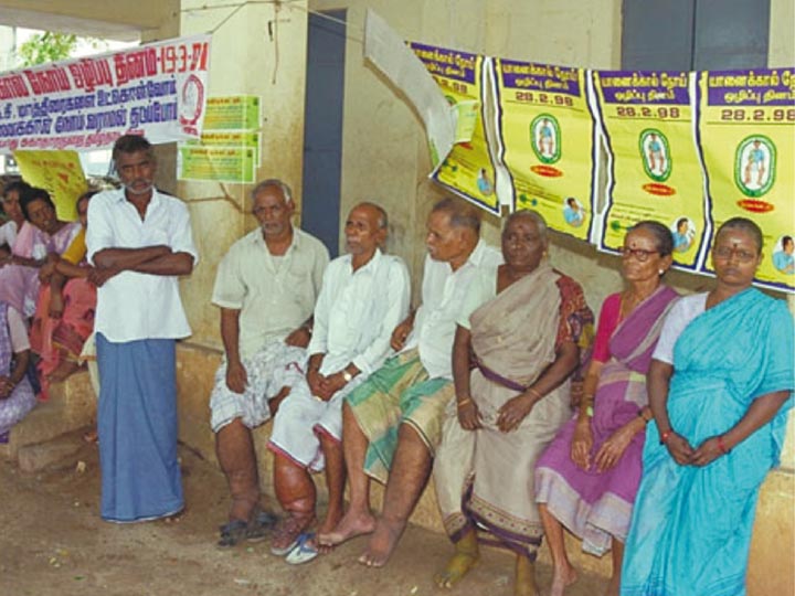
Figure 2: ‘Leg’ clinic in which immobility, the effects of gravitational stasis, and rare taking of deep breaths are featured.
Skin hygiene and moisturisation
As emphasised by Matts, by what I have called the Matts’ Hypothesis5, washing the skin with 3% glycerine19 (or using any locally preferred emollient) reduces transepidermal water loss, providing a very effective way of reversing a major contributor to lymphoedema. The epidermis reacts to moisturisation by switching off the repair response, and preventing cytokine production that activates dermal vasculature. I always emphasise switching off the repair mode of the epidermis, sealing entry points for bacteria and soil irritants, and moisturising what is often a desiccated skin surface. It should be applied to all the skin, especially if it is scaly and erythematous. I do not discourage local traditional practices that include herbal washes, but I try to put a stop to scarification as it adds to local inflammation and infection. It should be remembered that the skin prefers to be acidic and that many soaps are very alkaline. A teaspoon of vinegar in a pint of water can be beneficial to overcome this.
Of course there are many other interventions for the treatment of oedema, but mostly they add cost. Some are essential, such as footwear in environments with irritant soil. In recent years the association between lymphoedema and obesity has been identified. This has led to further research and the finding that adipose tissue is stimulated by lymphoedema, and its removal by liposuction helps in control of lymphoedema20. At the Institute of Applied Dermatology in Kerala, the Integrated medical system includes appropriate dieting.
Bandages and hosiery
Bandages and hosiery are the basis of most biomedical therapy (Figure 3). Manual lymphatic drainage (MLD) by a skilled practitioner, and skilled bandaging using modern varieties of short-stretch bandages, are unquestionably best practice. However, in resource-poor countries, these are not easily applied nor affordable. Having spent many days teaching patients and their families such best practice, I am invariably disappointed when, on visitation months later, I see how the bandages are applied: loose around the foot and constricting just below the knee. In resource-poor regions, I find bandaging skills so poor with the available material, that I do not insist on anything that is unaffordable. Notable is the fact that their lymphoedema, in spite of ineffective bandages, is improving. The reason for this is that the patients are moving more and are regularly taking care of their skin, including hygiene and moisture interventions. They are thus reducing lymphatic overload caused by inflammation and venous gravitational effects.
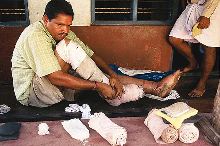
Figure 3: Some patients need many bandages, but have only simple cotton bandages worn out by washing. The effectiveness of their bandaging is questionable.
Conclusion
In the management of oedema and lymphoedema, biomedical practice has focused on MLD, bandages and hosiery. Low-cost, self-help interventions such as limb elevation, body movement, breathing, and weight control should be considered and promoted. Skin care strategies including hygiene and the application of emollients, can switch off the repair response, and prevent cytokine production that activates dermal vasculature, contributing to oedema formation. This discussion paper has highlighted simple but effective interventions which should be considered and implemented in a wide range of health care settings and environments including low-resource communities.
Author(s)
Terence J Ryan
DM, FRCP
Emeritus Professor of Dermatology
Oxford University, Oxford, UK
References
- Myers MB, Cherry G, Heimburger S et al. The effect of edema and external pressure on wound healing. Arch Surg 1967;94:218–222.
- Ryan TJ. The first commandment: Oil it. An appreciation of the science underlying water and emollients for skin care. J Community Dermatology 2004;1:3–5.
- Rawlings AV, Matts PJ. Stratum corneum moisturization at the molecular level: an update in relation to the dry skin cycle. J Invest Dermatol 2005;124(6):1099–1102.
- Ryan TJ. The skin as a barrier: What does it mean when it fails when lymphoedema is present. J Lymphoedema 2013;8(1):6.
- Ryan TJ. Matts’ Hypothesis: How simple strategies can lead to better outcomes. J Lymphoedema 2016;11:41–43.
- Dreyer G, Addiss D, Dreyer P, Noroes J. Basic Lymphoedema Management: Treatment and Prevention of Problems Associated with Lymphatic Filariasis. NH, USA: Hollis Publishing Co, 2002, 11.
- Dreyer G, Addiss D, Gadelha P et al. Interdigital skin lesions of the lower limbs among patients with lymphoedema in an area endemic for bancroftian filariasis. Trop Med Internat Health 2006;11:1475–8.
- Casley-Smith JR, Clodius L, Piller NB. Tissue changes in chronic experimental lymphoedema in dogs. Lymphology 1980;13:130–41.
- Belgrado JP, Vandermeeren L, Vankerckhove S et al. Deep Infrared imaging to identify venous impairment after breast cancer surgery. Eur J Lymphology and Related Problems 2015;26:20.
- Bates DO, Levick JR, Mortimer PS. Starling pressures in the human arm and their alterations in postmastectomy oedema. J Physiol 1994;477(pt.2):355–363.
- Moffatt CJ, Franks PJ, Doherty DC, et al. Lymphoedema: an underestimated health problem. QJM 2003;96:731–738.
- Narahari SR, Ryan TJ, Bose KS, Prasanna KR, Aggithata M. Integrating modern dermatology and Ayurveda in the treatment of vitiligo and lymphoedema in India. Int J Derm 2011;50:310–334.
- Ryan TJ, Narahari SR. Reporting an Alliance using an integrative approach to the management of Lymphoedema in India. Int J Lower Extrem Wounds 2012;11:5–9.
- Narahari SR, Bose KS, Aggithaya MG, Swamy GK, Ryan TJ, Unnikrishnan B, Washington RG, Rao BPS, Rajagopala S, Manjula K, Vandana U, Thaivalath TA, Rojith M, Shanappa Y. Shefuvan S and M (2013) Community level morbidity control of lymphoedema using self care and integrative treatment in two Lymphatic filariasis endemic districts of South India: a non randomized interventional study. Trans R Soc Trop Med Hyg 2013;9:566–577.
- Narahari SR, Ryan TJ, Aggithaya MG. How does yoga work in lymphoedema? J Yoga Phys Ther 2013;3:135.
- Aggithaya MG, Narahari SR, Ryan TJ. Yoga for correction of lymphedema’s impairment of gait as an adjunct to lymphatic drainage: A pilot observational study. Int J Yoga 2015;8:54–61.
- Narahari SR, Guruprasad M, Therme L, Bose KS, Ryan TJ. Yoga protocol for treatment of breast cancer-related lymphoedema. Int J Yoga 2016;9:145–55.
- Narahari SR, Aggithaya MG, Thermo L Bose KS, Ryan TJ. Response to comment on the article, Yoga protocol for treatment of breast cancer-related Lymphoedema. Int J Yoga 2017;10:52–4.
- Brooks J. An RCT to Determine an Effective Skin Regime Aimed at Improving Skin Barrier Function and Quality of Life in Those with Podoconiosis in Ethiopia. Unpublished Phd, University of Hull, 2016.
- Brorson, H, Svensson, H, Norrgren, K, Thorsson O. Liposuction reduces arm lymphoedema without significantly altering the already impaired transport. Lymphology 1998;32:156–172.

