

Volume 26 Number 4
Case report: Necrotising fasciitis after removing the intrauterine device
Birgul Özkaya, Asiye Gul, Ayşegul Kuçuk and Hale Tosun
Keywords wound., Care, necrotising fasciitis, vacuum-assisted closure
Abstract
Necrotising fasciitis is a rapidly progressive, destructive soft tissue infection that mainly involves fascia and subcutaneous tissues. Rapidly spread necrosis in tissues is often caused by systemic sepsis, toxic shock syndrome, multiorgan failure and thrombosis in the subcutaneous vasculature. Necrotising fasciitis can be seen in all the anatomical regions of the body; the extremities and perineum are frequently affected. In this case report, we emphasised the importance of nursing care of a necrotising fasciitis patient who has been treated for a long time with aggressive surgical treatment. Vacuum-assisted closure application is a non-invasive method with controlled and localised negative pressure on the wound to accelerate healing in acute/chronic wounds. Necrotising fasciitis can be successfully treated with early diagnosis, adequate debridement, and appropriate antibiotic therapy. A multidisciplinary approach is necessary for the comprehensive care of these patients.
Introduction
Necrotising fasciitis (NF), commonly known as flesh-eating disease, is caused by a bacterial infection that results in the death of the body's soft tissue1. Hippocrates described this condition as “Diffused erysipelas caused by trivial accidents, [where] flesh, sinews, and bones fell away in large quantities, [leading to] death in many cases”2. NF is a rare and lethal bacterial infection characterised by progressive necrosis of the skin, subcutaneous tissue, and fascia3-5. NF initially begins as a localised infection, with only mild redness and swelling on the skin6. Then, with the synergistic effects of microorganisms and some risk factors, it progresses rapidly within 3–4 days and causes bulla and necrosis of the skin5,6. Pain may occur hours before infection4. Pain is one of the most sensitive symptoms, seen in almost all patients and disproportionate to the lesion5. Rapidly spread necrosis in tissues is often caused by systemic sepsis, toxic shock syndrome and multiorgan failure and thrombosis in the subcutaneous vasculature4,7. Regardless of the causative pathogen, early diagnosis is difficult because there is no deep involvement of the necrotic area during the initial period3.
Although NF can be seen in all ages and both genders, it occurs more frequently in males and aged 50–60 years4. The incidence is 0.4 in 100,000 cases3,5. Mortality is reported to be 6–76%5. The principle of surgical management is immediate and extensive radical debridement of necrotic tissues and fasciotomy1,2. The delay of the first surgical debridement may increase the mortality3.
There are some risk factors that facilitate for NF: immune system disease, ageing, diabetes, obesity, debility, alcohol dependence, intravenous drug use, peripheral vascular disease, smoking, cancer, advanced liver and kidney diseases, trauma or surgery4,5,8. In the literature, diabetes has been reported in 60% of patients5,6. Although there are many identified risk factors, half of the cases occur in healthy individuals5.
NF can be seen in all the anatomical regions of the body4. However, the most common sites of infection are the lower extremities (28%), perineum (21%), trunk (18%), and head or neck (5%)2. NF is usually characterised by minor injuries such as surgical incision, insect insertion, incision, abrasion, contusion, injection, skin ulcer, perirectal abscess, incarcerated hernia, burns, splintering, birth and penetrant trauma5. Extremities often develop after trauma, injection, or insect bites, and frequently in the abdominal region as an operative complication7.
After the description by Jean Alfred Fournier in 1883, it was called “Fournier's gangrene” when external genital organs and perineum were affected5. Fournier’s gangrene typically begins in the labium region in women and in the scrotum region in men and spreads rapidly to the perineal, gluteal, and abdominal regions4. Other anorectal aetiologic factors include perianal abscess, diverticulum perforation, cancer, rectal perforation, acute appendicitis perforation, anal dilatation, anal fistula, and strangulated inguinal hernia7. The common aetiologic factors among urogenital lesions are urethral stenosis and instrument use8. If NF occurs after a known aetiology, it is classified as secondary NF. In 45% of cases, no definite cause can be detected, and it is called primary or idiopathic NF5.
Depending on the body area and underlying predisposing factors developed by the infection, different types of microorganisms are isolated6. The most frequently isolated bacteria are Streptococcus, Staphylococcus and Escherichia species9.
Amputation in limb involvement can be life-saving. However, there is a higher mortality rate because amputation cannot occur in the trunk and perineal involvement5. In cases where the perineum is affected, early diagnosis, early debridement, and close monitoring are the only options to reduce mortality rates3. It has been shown that a 12-hour delay in surgical debridement leads to an increase in the incidence of septic shock and acute renal failure and a 24-hour delay in surgical debridement increases mortality6.
In this case report, we aimed to emphasise the importance of the care and treatment of a patient who developed uterine rupture and NF during removal of an intrauterine device.
Ethical consideration
The study was carried out according to the principles of the Declaration of Helsinki. Verbal and written permission was obtained from the patient.
Case report
History
A 39-year-old female patient lives with her husband and four children. The uterus rupture developed while the intrauterine device was removed from the patient. The patient, who was sent home without noticing the rupture, began complaining of severe abdominal pain, fever, nausea, vomiting and incontinence after one month. On clinical examination, the patient was found to have lethargy and fever, with a temperature of 38 °C. The patient was admitted to our emergency clinic. Necrotic tissue was found inside the anterior abdominal wall (20x10 cm in width). Blood investigation showed leukocytosis.
Treatment
Emergency surgery was performed after examination and evaluation. A colostomy was opened due to sigmoid colon perforation in the patient and Vacuum-Assisted Closure® (VAC) was applied to the abdomen because the frontal wall of the abdomen was necrotic. Culture samples from necrotic tissue and blood culture were taken and sent to the microbiology laboratory. Intravenous fluid therapy was initiated by opening vascular access. Until culture results were available, broad-spectrum intravenous antibiotics were started. The patient was taken to the operating room. Around the wound, black polyurethane sponges were cut with scissors in the appropriate sizes. Empty spaces in hollow wounds were whipped with black polyurethane sponges. The wound areas were covered with adhesive, semi-permeable covers. A small hole was made through the closures to place the TRAC™ (Therapeutic Regulated Accurate Care) pad and the VAC device was connected to the wound. Dressing changes were made in 48–72 hours according to the condition of the wound. During the dressings, superficial debridement was made when needed (Figure 1). The patient and her family were informed about the gravity of their condition. The VAC application was performed until the wound was ready to surgically close.
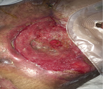
Figure 1: Application of debridement to the wound.
During the third VAC, the perforation of the small intestine was observed, and two ostomies were made. On the 15th day, the general condition of the patient was deteriorated, and the signs of sepsis were seen. The patient was transferred to the critical care unit. In order to correct the patient’s haemodynamic instability, antibiotic treatment, hypoalbuminaemia treatment, and blood supplements were applied. After three days in the critical care unit, when her haemodynamic parameters were stable, she was taken to the general surgery clinic again. Triple antibiotherapy was started for wound infection treatment. The application of VAC to the abdominal region continued for a month and VAC was changed every three days.
Prognosis
The VAC application was finished because of wound infection to a minimal level, and reduction of wound area measurements (7X12). The patient was discharged well after 90 days (Figure 2).
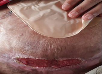
Figure 2: On the 90th day of wound healing.
Discussion
NF is a rare but rapidly progressive and potentially fatal infection10. The care for a patient with NF is multidimensional and multidisciplinary, requiring nursing interventions11. In this case report, a 39-year-old female patient developed uterine rupture and NF after intrauterine device removal. This infection can occur at any age or in any location and is not specific to gender11. Older age (>60) is a risk factor for NF10. Özgenel et al.7 retrospectively examined 30 cases of NF (4 female and 26 male) for a 5-year period; the mean age was 55 years. Bozkurt et al.12 reported a 22-year-old male patient with NF due to razor self-mutilation in the forearm. Vayvada et al.5 examined a total of 68 patients who had NF in a 10-year period and found that 76.4% (n=52) of patients had risk factors such as diabetes, smoking, obesity, and the use of corticosteroids. The most common predisposing factor in the study of Demir and Kunt13 was diabetes mellitus (71.4%), followed by peripheral vascular disease (21.4%) and malignancy (14.3%), respectively. No predisposing factor was detected in our case, but most patients have a history of trauma or may have a history of surgical or penetrating injury10. In this case, weakening of the immunity system after uterine rupture may also have contributed.
NF is difficult to diagnose because the symptoms are many and varied13. Early diagnosis and aggressive surgical treatment reduce mortality risk12. The basis of the treatment is an early diagnosis, broad-spectrum antibiotic therapy, debridement reaching intact tissues with no necrotic tissue left behind, regulating liquid-electrolyte balance, adequate oxygenation of the infected area and sufficient nutritional support and analgesia supply4. Sepsis is a frequent complication of NF. Bacterial cultures and antibiotic sensitivity can help in the treatment process14. Laboratory findings include leukocytosis with an elevated number of neutrophils11. The usual infection control precautions such as hand hygiene are important. The patient should be in a private room and under contact isolation.
The patient with NF has many psychological needs11. Our patient had two ostomies, which resulted in impaired body image; her husband couldn’t accept the stoma. Body image refers to a subjective concept of the physical appearance of the person based on her own self-observation and the reaction of others15. Changes in physical appearance, function and body integrity are typically central to disease experience and medical treatment16. Our patient and husband were informed that these changes in her body were temporary and that the stoma would be closed after a while.
Third-degree pressure injury developed due to immobility and reluctance to meet self-care needs. A deterioration of skin integrity, lack of knowledge, malnutrition due to oral intake impairment, weight loss and prolonged sleep were observed. Replacing the patient’s nutritional requirements is necessary to promote wound healing11. Total parenteral nutrition and enteral nutrition started for weight loss. In these patients, arterial blood pressure and central venous pressure, and daily urine output should be monitored. In addition, correction of metabolic acidosis and electrolyte imbalance is one of the most important interventions14.
The timing of surgery is extremely important and is predicated on prompt diagnosis14, and VAC application; it is a non-invasive method with controlled and localised negative pressure on the wound to accelerate healing in acute and chronic wounds13. This therapy helps remove infectious materials and other fluids from the wound. VAC is typically applied to the wound after debridement when clear edges have been obtained11. The VAC system consists of polyurethane and polyvinyl alcohol sponges to fill the wound, semi-permeable caps with adhesive to cover the wound, a device that provides device connection with the wound, a pressure regulator, a collecting container and a device that produces a negative pressure8. It both reduces bacterial burden by removing from the environment the excretion of the bacteria and allows the capillary to overcome the pressure and bring more blood, thus more oxygen and accompanying growth factors5. The VAC is changed every two to three days. By the VAC applied to the wound in our case, the wound surface was reduced at the end of one month and the VAC was removed. VAC is now becoming an alternative treatment option that is becoming increasingly widespread in acute and chronic wound treatment.
During the VAC application, superficial debridement was applied when necessary. Early surgical debridement is necessary to remove all necrotic tissue and prevent the spread of the infection11. Without surgical intervention, mortality approaches 100%10. Özgenel et al.7 found that 96.6% of patients required more than one debridement. In the study of Demir and Kunt13, they reported that all patients had to have received wound care at least twice a day. Pain is an important issue in the patient with NF11. For this reason, pain control was provided by appropriate painkillers before the procedure. Two ostomies were performed in this patient due to small intestinal perforation. Firstly, patients and their relatives were informed. Her husband was taught stoma care. When managing these patients, intensive care facilities should be provided and long-term follow-up with the patient and treatment of complications should be discussed and planned.
Conclusion
NF is a form of infection that affects soft tissues and fascia is rapidly progressing and life-threatening. NF can be successfully treated with early diagnosis, adequate debridement, and appropriate antibiotic therapy. A multidisciplinary approach is necessary for the comprehensive care of these patients. For this reason, a multidisciplinary approach is needed in the diagnosis and treatment of NF cases.
Authorship and Acknowledgements
The manuscript has been read and approved by all authors and all authors agree to the submission of the manuscript.
All authors must have agreed on the final version of the paper and must meet at least one of the recommendations in the International Council of Medical Journal Editors criteria.
Conflict of Interest
The authors declare that there is no any conflict of interest for this work.
Funding
The authors received no funding for this study.
Author(s)
Birgul Özkaya
MSc, Staff Nurse
Bakırkoy Dr Sadi Konuk Training and Research Hospital, General Surgery Clinic
İstanbul, Türkiye
Asiye Gul*
PhD, Associate Professor
İstanbul Kültür University, Faculty of Health Science
İstanbul Kültür Üniversitesi Şirinevler Yerleşkesi
İstanbul, Türkiye
Email asiyegul2003@yahoo.com
Ayşegul Kuçuk
Staff Nurse
Bakırkoy Dr Sadi Konuk Training and Research Hospital, General Surgery Clinic
İstanbul, Türkiye
Hale Tosun
PhD, Assistant Professor
University of Health Science, Faculty of Health Science
İstanbul, Türkiye
* Corresponding author
References
- Ali AH, Salahuddin Z, Ismail H, Sofi AIM, Mohamad I. Debridement of facial necrotizing fasciitis via bicoronal flap. Egyptian Journal of Ear, Nose, Throat and Allied Sciences 2017;18:287–289.
- Lancerotto L, Tocco I, Salmaso R, Vindigni V, Bassetto F. Necrotizing fasciitis: classification, diagnosis, and management. J Trauma 2017;72(3):560–566.
- Altintop İ, Baykan H, Tatlı M, Yurtseven A, Ay Altıntop Y. Fatal necrotizing fasciitis: a case report. Bozok Med J 2016;6(4):68–71.
- Uluğ M, Çelen MK, Geyik MF, Hoşoğlu S, Ayaz C. A case of necrotizing fasciitis due to intramuscular injection and review of the literature. Klimik Dergisi 2009;22(3):103–5.
- Vayvada H, Demirdöver C, Menderes A, Karaca C. Necrotizing fasciitis: diagnosis, treatment and review of the literature. Ulus Travma Acil Cerrahi Derg 2012;18(6):507–513.
- Akbaş T, Bilgin H, Aykut UE, Karakurt S. Necrotizing fasciitis: delayed surgical intervention increases mortality. Düzce Üniversitesi Sağlık Bilimleri Enstitüsü Dergisi 2015;5(3):84–88.
- Özgenel GY, Akın A, Kahveci R, Özbek S, Özcan M. Clinical evaluation and treatment results of 30 patients with necrotizing fasciitis. Turkish Journal of Trauma & Emergency Surgery 2004;10(2):110–114.
- Ilhan M, Gemici E, Yanar F, Kapan S, Alış H. 5-year single center experience of necrotizing fasciitis treatment approaches. Medical Journal of Bakırkoy 2014;10(2):70–74.
- Singh A, Ahmed K, Aydin A, Khan MS, Dasgupta P. Fournier’s gangrene. A clinical review. Arch Ital Urol Androl 2016;88(3):157–164.
- Puvanendran R, Huey JCM, Pasupathy S. Necrotizing fasciitis. Can Fam Physician 2009;55:981–987.
- Ellis P, Murray TA. Caring for the patient with skin disorders. In KS Osborn, CE Wraa, AB Watson, Editors. Medical-Surgical Nursing Preparation for Practice. Boston: Pearson Education; 2010, pp. 2149–2191.
- Bozkurt M, Zor F, Külahçı Y, Benlier E. Necrotizing fasciitis in forearm: Case report and literature review. Cerrahpaşa J Med 2006;37:17–19.
- Demir CY, Kunt A. Necrotizing soft tissue infections: Our clinical results. İnönü Üniversitesi Tıp Fakültesi Dergisi 2003;10(4):187–190.
- Liu Y, Guo K, Sun J. Learning from clinical experience with necrotizing fasciitis: treatment and management. Adv Skin Wound Care 2017;30(11):486–493.
- Martin JB. The development of ideal body image perceptions in the United States. Nutr Today 2010;45(3):98–110.
- Bolton MA, Lobben I, Stern TA. The impact of body image on patient care. Prim Care Companion J Clin Psychiatry. 2010;12(2):PCC.10r00947.

